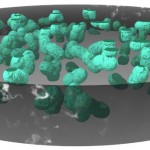
This post is about how to visualize non-filamentous cyanobacteria by taking the genus Aphanotheca as an example. Colonies of Aphanotheca show a rather simple morphology: a bunch of individual cells, clearly separated from each other is embedded in a globular gel-like matrix.
- Aphanotheca
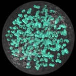
The next image simulates microscopy after ink-staining. (Incident light from below the object is absorbed by the ink but scattered by the gel-like matrix).
- Aphanotheca
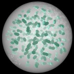
Now I add some light from above to improve the image of the individual cells.
- Aphanotheca
Finally, it is possible, at least when using ray-tracing, to give the gel-like matrix a darker colour.
- Aphanotheca
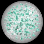
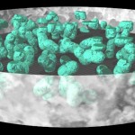
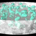
One of the great advantages of ray-tracing when compared to microscopy is the ease of preparing sections 😉 Here I am doing rather thick ones, but there is no problem doing ultrathin sections as well. (Well in this case there is no point as well…)
- Aphanotheca
I am using several ways of illumination below.
- Aphanotheca
- Aphanotheca
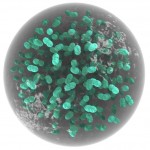
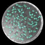
And finally, how such a section would appear in bright field microscopy.
In the next post on cyanobacteria I will apply these ways of visualization to the various forms of non-filamentous cyanobacteria.
Further reading:
Exploring cyanobacterial diversity in Antarctica blog
Did Cyanobacteria Produce Oxygen Oceanic Oases?

Hi Thomas,
This is a great blog! I received my Ph.D. working with a soil bacterium (Azospirillum), so I can appreciate your research. Great illustrations as well (something I use on my blog). Please keep up the great work!
Hi Matt,
sorry for this late reply, but I had received such a huge amount of spam to this blog… And then I had less and less time, so I paused it for a while. It seems I am going to reanimate it soon, but in another form and closely connected to my youtube account (https://www.youtube.com/channel/UCittg4tLzmxR_xsgWlwXpxA).
Thomas